-
×
 The Brigham Update In Hospital Medicine 2022
1 × $40.00
 -
×
 SCCM Multiprofessional Critical Care Review Adult 2024
1 × $60.00
 -
×
 BMJ Learning Online (1-year Subscription)
1 × $20.00
 -
×
 USMLE® Easy Subscription (Including Step 1, 2, 3) (1-year Subscription)
1 × $20.00
 -
×
 Web of Science (1-year Subscription)
1 × $20.00
 -
×
 ASAM Virtual Review Course in Addiction Medicine 2023 (Videos)
1 × $55.00
 -
×
 2023 ASCO Annual Meeting
1 × $55.00
 -
×
 Apollo Clinical Research Conclave 2022
1 × $10.00
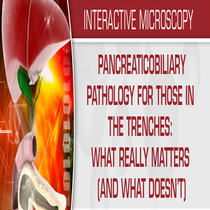
Pancreaticobiliary Pathology for Those in the Trenches What Really Matters (and What Doesn’t) 2020 (VIDEOS)
$30.00
Pancreaticobiliary Pathology for Those in the Trenches What Really Matters (and What Doesn’t) 2020 (CME VIDEOS) Every general surgical pathology practice routinely receives gallbladder specimens, yet many pathologists lack sufficient expertise in gallbladder pathology to feel comfortable when confronted with unusual patterns of inflammation such as vasculitis, parasitic infestation, and immune-mediated conditions.
Samples for Courses Can be found here : Free Samples Here!
Categories: Courses, Obstetrics & Gynecology COURSES, Pathology courses
Tags: Obstetrics & Gynecology COURSES, Pathology
Pancreaticobiliary Pathology for Those in the Trenches What Really Matters (and What Doesn’t) 2020 ( VIDEOS)
Course Description
- Pancreaticobiliary Pathology for Those in the Trenches What Really Matters (and What Doesn’t) 2020 (CME VIDEOS) Every general surgical pathology practice routinely receives gallbladder specimens, yet many pathologists lack sufficient expertise in gallbladder pathology to feel comfortable when confronted with unusual patterns of inflammation such as vasculitis, parasitic infestation, and immune-mediated conditions. Gallbladder neoplasms are uncommon and often discovered at the time of histologic examination, provoking angst among surgeons and pathologists who infrequently encounter such cases and may not be aware of updated terminology and staging issues. Widespread use of cross-sectional imaging in the evaluation of patients with abdominal symptoms has led to increased numbers of incidentally discovered pancreaticobiliary lesions. Many of these are initially evaluated with fine-needle aspiration biopsy (FNAB) or limited tissue biopsy samples. As a result, pathologists are often faced with cytology, biopsy and resection specimens that feature disorders relatively uncommon to their practices. They may be required to deal with such specimens under pressure, such as in the frozen section laboratory where immunohistochemical stains and other ancillary tools are not available. This course is intended to provide learners with practical information and diagnostic pearls that may aid them in their day-to-day practices.
Target Audience
Practicing academic and community pathologists, and pathologists-in-training
Learning Objectives
Upon completion of this educational activity, learners will be able to:
- Discriminate between carcinoma and its potential mimics in frozen sections and limited biopsy material
- Understand the classification and differential diagnoses of cystic and solid intraductal lesions
- Recognize and classify important inflammatory conditions of the pancreaticobiliary tree
- Develop an algorithmic approach to differential diagnosis of solid pancreaticobiliary tumors
- Generate a differential diagnosis for lesions encountered in cytology specimens
- Continuing Medical Education and Continuing Certification
The United States and Canadian Academy of Pathology is accredited by the Accreditation Council for Continuing Medical Education (ACCME) to provide continuing medical education for physicians.
The United States and Canadian Academy of Pathology designates this enduring material for a maximum of 11.25 AMA PRA Category 1 CreditsTM. Physicians should claim only the credit commensurate with the extent of their participation in the activity.
USCAP is approved by the American Board of Pathology (ABPath) to offer Self-Assessment credits (SAMs) and Lifelong Learning (Part II) credit for the purpose of meeting the ABPath requirements for Continuing Certification (CC). Registrants must take and pass the post-test in order to claim SAMs credit. Physicians can earn a maximum of 11.25 SAM/Part II credit hours.
Disclosures
- The faculty, committee members, and staff who are in position to control the content of this activity are required to disclose to USCAP and to learners any relevant financial relationship(s) of the individual or spouse/partner that have occurred within the last 12 months with any commercial interest(s) whose products or services are related to the CME content. USCAP has reviewed all disclosures and resolved or managed all identified conflicts of interest, as applicable.
- The following faculty reported no relevant financial relationships: Wendy Frankel, MD, Jose Jessurun, MD, Martha Pitman, MD and Rhonda K. Yantiss, MD
- The following IM Coordinator who planned and reviewed content for this activity reported no relevant financial relationships: Steven D. Billings, MD
- USCAP staff associated with the development of content for this activity reported no relevant financial relationships.
Topics/Speaker:
- – Beyond Chronic Cholecystitis Non-Neoplastic Diseases of the Gallbladder and Bile Ducts
- – Challenges in the Diagnosis of Pancreas Adenocarcinoma
- – How to Sign Out Cytology of Pancreatic Cysts
- – Pancreas Frozen Sections What the Surgeon Needs to Know and How to Stay Out of Trouble
- – Pancreaticobiliary Cysts and Things That Look Like Them
- – PDAC or Not-That Is the Question- From FNA to BDB
- – Solid Pancreatic Tumors WHO Made All This So Complicated
- – Unfortunate Outcomes of Chronic Inflammation Neoplasms of The Gallbladder, Bile Ducts, and Ampulla
Original release date: March 27, 2020
Access to this course expires on: January 26, 2023

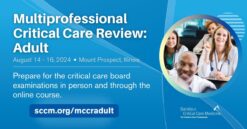 SCCM Multiprofessional Critical Care Review Adult 2024
  BMJ Learning Online (1-year Subscription)
  USMLE® Easy Subscription (Including Step 1, 2, 3) (1-year Subscription)
  Web of Science (1-year Subscription)
  ASAM Virtual Review Course in Addiction Medicine 2023 (Videos)
  2023 ASCO Annual Meeting
  Apollo Clinical Research Conclave 2022